
Sonosa Medical Publishes Ultrasound Against Sleep Endoscopy in The Laryngoscope
In a trial run alongside the procedure it aims to replace, machine learning models reading airway ultrasound reproduced the endoscopist's findings at four of five airway sites.
MDC Studio portfolio company Sonosa Medical has published results in The Laryngoscope comparing airway ultrasound directly against drug-induced sleep endoscopy, the procedure clinicians currently rely on to decide how to treat obstructive sleep apnea. It is the first study to compare ultrasound characteristics with sleep endoscopy outcomes using the standard VOTE criteria.
The comparison was made under the fairest possible conditions: rather than testing the two methods on different patients on different days, ultrasound was recorded during the endoscopy itself. Twenty adults undergoing clinical sleep endoscopy at Ascension Saint Agnes Hospital in Baltimore were imaged submentally while awake and again while sedated, with the endoscope in place. Both methods therefore observed the same airway, in the same patient, at the same depth of sedation.
What the trial found
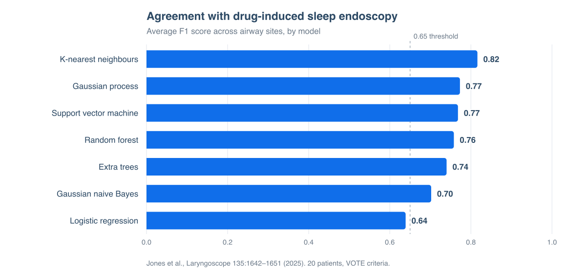
Three independent clinical reviewers graded the endoscopy video for collapse at each VOTE site — velum, oropharynx, tongue base, and epiglottis. Machine learning models were then trained to predict those grades from ultrasound measurements alone. The models reproduced the endoscopists' binary scores at four of the five sites, with the strongest result at lateral velum collapse, where agreement reached an F1 score of 0.93 averaged across models. Across all sites, k-nearest neighbours averaged 0.82.
A parallel regression analysis, predicting the degree of collapse on a continuous scale rather than a yes-or-no grade, explained between 53% and 82% of the variation depending on the site, with the tongue base the best modeled at 82%.
Drug-induced sleep endoscopy is the reference standard because it shows the airway collapsing in real time. But it has to be performed in an operating room, with an anesthesiologist, operating room staff, and operating room time, which makes it among the most expensive steps in the whole sleep apnea pathway. It also carries the risk of sedating a patient whose defining problem is that their airway closes.
And what it observes is not sleep. Propofol sedation produces a drug-induced state that only approximates natural sleep, and the depth of that sedation itself changes how much the airway collapses — a known limitation of the procedure, and one the study acknowledges in its own results. Grading is subjective as well: the paper notes variation between its three reviewers. Ultrasound, by contrast, is inexpensive, uses no ionizing radiation, and needs no sedation at all.
What the trial does not yet show
The authors are direct about the limits of a pilot at this scale. Twenty patients is a small dataset for machine learning, and while permutation testing supported the models for lateral collapse at the velum and oropharynx (p < 0.02), the anterior-posterior categories did not reach statistical significance. There was no control group without sleep apnea, so the study cannot show that ultrasound diagnoses the condition — only that it tracks the collapse patterns endoscopy reveals. Holding a consistent imaging plane by hand during breathing proved difficult and contributed to measurement variability. Larger studies are needed for accuracy and generalizability.
That hand-held variability is the problem Sonosa's wearable is built to remove, and it closes the wider gap as well. ApneaView images the airway automatically through the night during natural sleep — no sedation, no operating room, no operator holding a probe — and it is designed to be worn at home, in the patient's own bed, rather than in a hospital. The study establishes what ultrasound can see of airway collapse; the wearable is what carries that capability out of the operating room.
The work was published as Ultrasound Predicts Drug-Induced Sleep Endoscopy Findings Using Machine Learning Models by Samuel E. Jones, Natalie Aw, Molly Acord, Sarah Miller, Danielle Sidelnikov, Sunny J. Haft, and Stephen M. Restaino, in The Laryngoscope 135:1642–1651 (2025), doi:10.1002/lary.31950. It was supported by Small Business Innovation Research grants to Sonosa Medical from DARPA and the National Institutes of Health. Sonosa Medical is a for-profit company seeking to commercialize this research; the senior author is a shareholder, and three co-authors were employees at the time of the work.
About Sonosa Medical
Sonosa Medical develops ApneaView, a wearable ultrasound system that images the upper airway during natural sleep at home to identify where obstruction occurs, information that today requires an endoscopy under sedation. The company was founded in the MDC Studio and is part of the MDC Studio portfolio. sonosamedical.com
About the MDC Studio
The MDC Studio is a MedTech startup studio in Baltimore, Maryland. It works with clinicians and engineers to turn ideas into working prototypes and into companies, providing engineering, business development, regulatory support, and shared laboratory and office space. The Studio is located in an Opportunity Zone at 300 West Pratt Street. Contact us for more information.